Many thanks to Hellequin GB for translating this amusing clip from Germany, and to Vlad Tepes and RAIR Foundation for the subtitling:
Video transcript:


Many thanks to Hellequin GB for translating this amusing clip from Germany, and to Vlad Tepes and RAIR Foundation for the subtitling:
Video transcript:
 Parties and other lockdown-violating gatherings were targeted for fines in Ontario, Quebec, Malta, the UK, and Jersey. Meanwhile, a man was kicked off a Southwest Airlines flight for not putting his mask back on between bites of food.
In other news, according to the latest polls, a majority of French people agree with the open letter from retired generals warning President Macron that civil war is imminent.
To see the headlines and the articles, click “Continue reading” below.
Thanks to Dean, DV, László, Reader from Chicago, SS, and all the other tipsters who sent these in.
Notice to tipsters: Please don’t submit extensive excerpts from articles that have been posted behind a subscription firewall, or are otherwise under copyright protection.
Caveat: Articles in the news feed are posted “as is”. Gates of Vienna cannot vouch for the authenticity or accuracy of the contents of any individual item posted here. I check each entry to make sure it is relatively interesting, not patently offensive, and at least superficially plausible. The link to the original is included with each item’s title. Further research and verification are left to the reader.

Hellequin GB has translated three articles from PolitikStube on migration-related news from Germany.
The first article reports on Germany’s front-runner status in the “asylum” race:
Germany leads the way in asylum applications in the first quarter
With over 26,000 asylum applications in the first quarter of this year, Germany is the front-runner in the EU. In contrast, Estonia accepted eleven and Hungary only eight asylum applications.
“The figures come from a confidential status report by the EU Commission on the current migration situation,” reports Welt am Sonntag and refers to previously unpublished figures from the EU asylum authority EASO (European Asylum Support Office).
It also emerges that, according to the document, 41% of applicants in Germany are Syrians, followed by Afghans (14%) and Iraqis (7%).
This outrage, according to which Germany receives by far the greatest number of asylum applications in Europe, must be ended immediately. In addition, incentives in the German asylum procedure — for example, excessively long procedures, access to the labor market before recognition, excessive social benefits, failed deportations — that primarily attract asylum seekers within the EU to Germany must be removed.
The second article concerns the abandonment of criminal proceedings investigating official corruption in the awarding of asylum status to applicants:
Criminal proceedings against the former head of the Bremen refugee office stopped
It all started in October 2017, when a forged (Asyl) Decision from the Bremen branch of the Federal Office for Migration and Refugees (BAMF [Bundesamt für Migration und Flüchtlinge]) in Hesse emerged. In the spring of 2018, the public prosecutor began to investigate. The suspicion: The former head of the migration authority, together with several lawyers, wrongly helped hundreds of asylum seekers to obtain positive decisions. The public prosecutor’s office was investigating violations of official secrecy, data falsification and taking advantage.
Now the Bremen regional court has ruled, and the proceedings against the former head of the Bremen BAMF branch office have been discontinued against payment of €10,000. In September 2019, the Bremen public prosecutor’s office initially listed 121 alleged crimes that the ex-head of the agency and two lawyers allegedly committed. One of the investigators’ central allegations was that they illegally prevented foreign clients of the lawyers from being deported or improved their residence status.
Uwe Witt, chairman of the AfD [Alternative für Deutschland, Alternative for Germany] parliamentary group in the Labor and Social Affairs Committee of the German Bundestag: “The Bremen example clearly shows how lawyers, senior officials and the judiciary work together to smuggle asylum seekers into our social system. The judgment of the regional court strongly reminds me of the saying “One crow does not prick the other’s eye.”
And who has to pay for it? The German taxpayer.
The final article concerns the German government’s questionable official statistics on the number of Muslims in Germany:

A couple of weeks ago I posted a translation about a family law judge in Weimar who prohibited students and teachers from wearing masks in school, and overturned other provisions required by official national Corona policy.
As might have been expected, it’s unwise for any public official to oppose the narrative about the Wuhan Coronavirus as handed down from on high: the public prosecutor in Erfurt has ordered a raid on the judge’s residence, and his phone and computer have been seized.
Many thanks to Hellequin GB for translating this article from RedaktionsNetzwerk Deutschland, which is closely associated with the SPD (Social Democratic Party):
House search of the Weimar family judge: Public prosecutor investigates for perversion of the law
A Weimar family judge had ruled against the mask requirement in schools and justified this with the child’s best interests.
The administrative court overturned its judgment.
Now the family judge is being investigated for perversion of the law — and his cell phone and laptop have been confiscated.
The controversial judgment of a family judge at the Weimar district court against the Corona mask requirement at two Thuringian schools has consequences. The public prosecutor’s office in Erfurt has initiated an investigation against the judge, said the spokesman for the authorities on Monday. There is an initial suspicion of perversion of the law. The question is whether the judge has exceeded his jurisdiction with his corresponding decision. The public prosecutor’s office had received several reports against the man.
In the course of the investigation, the judge’s house was searched and his cell phone and laptop were confiscated. “This accusation is objectively and subjectively completely untenable,” said his defense lawyer, the renowned Hamburg lawyer Gerhard Strate, to the RedaktionsNetzwerk Deutschland (RND). “What the judge wrote may be disputed with good arguments, but it is carefully justified and by no means absurd. Only the judiciary can raise the accusation of perversion of the law if it anticipates the expected nuisance by the executive in advance obedience.”
Richter had stated that schools would play no role in the course of the pandemic According to the public prosecutor’s office, there are indications that the family judge arbitrarily assumed his jurisdiction, even though it was an administrative matter. The judge issued a temporary injunction at the beginning of April that children at two schools in Weimar would not have to wear Corona masks in class, contrary to the current hygiene concept of the Ministry of Education.
In his decision, the family judge relied on three reports that deny the effectiveness of various Corona protective measures. In addition, the judge had said schools played no role in the course of the pandemic.
The Weimar Administrative Court, on the other hand, recently declared the mask requirement at Thuringian schools to be permissible in class in an urgent procedure. The administrative judges classified the decision of the family judge as “obviously unlawful”. The family court has no authority to issue orders to authorities and their representatives. There is no legal basis for such a competence arrangement, it said.
 Chicago health authorities are planning to implement a “vax pass” system that will allow citizens to attend concerts and other public events if they have been vaccinated against COVID-19. Meanwhile, recent reports suggest that New York City lost $1.2 billion in tax revenues because of the collapse of the tourism industry after the “pandemic” hit.
In other news, three people were hospitalized for hypothermia after about 100 migrants attempted to swim from Morocco to the Spanish North African enclave of Ceuta.
To see the headlines and the articles, click “Continue reading” below.
Thanks to Fjordman, JM, PM, Reader from Chicago, SS, and all the other tipsters who sent these in.
Notice to tipsters: Please don’t submit extensive excerpts from articles that have been posted behind a subscription firewall, or are otherwise under copyright protection.
Caveat: Articles in the news feed are posted “as is”. Gates of Vienna cannot vouch for the authenticity or accuracy of the contents of any individual item posted here. I check each entry to make sure it is relatively interesting, not patently offensive, and at least superficially plausible. The link to the original is included with each item’s title. Further research and verification are left to the reader.

The N-VA (Nieuw-Vlaamse Alliantie, New Flemish Alliance) is a Flemish separatist party in Belgium. In the following video you’ll see a member of parliament for the N-VA, Mathias Vanden Borre, trying to escape the efforts of a group of “youths” in Brussels who seek to culturally enrich him. The video ends just as the beating begins.
Even though Mr. Vanden Borre is a member of a party that wants Dutch to be the sole official language in Flanders, for some reason he is speaking French when making the recording on his cell phone.
Many thanks to MissPiggy for the translation, and to Vlad Tepes for the subtitling:
Thanks to Gary Fouse for translating this Dutch-language article from Het Laatste Nieuws about the attack on Mathias Vanden Borre:
MP films how Brussels youths attack him: “Stomped, beaten and almost robbed”
[Photo caption: Brussels MP Mathias Vanden Borre (N-VA) suddenly surrounded and receives a couple of heavy blows]
Brussels MP Mathias Vanden Borre (N-VA) has made a film in which can be seen how he was attacked by a number of youths. “I was attacked, stomped, beaten, and almost robbed,” Vanden Borre tells HLN. The MP was able to flee but sustained injuries. The police have confirmed the story and are conducting an investigation.
The events occurred around 5pm in Brussels in the Essegem neighborhood, on the border of Jette and Laken. “I was with my wife and my 7-day-old son visiting a photographer. While my wife and my son were still inside, I went out for a walk around the block,” said Vanden Borre to HLN. The N-VA member of Parliament knows the area because he himself lives nearby.
“Suddenly six or seven young guys came walking behind me. They began yelling at me to intimidate me. I grabbed my cell phone and began to film. Then they came closer. I first got a kick in my side, then a blow on my jaw. One of them tried to steal my cell phone. Then I ran.”
Vanden Borre was able to escape, but the gang ran after him. “Then I ran another 500 meters and called the police.” They were at the scene within a few minutes with several teams, according to the MP.
Ringing in the ears
“My jaw is swollen, and I suffer from ringing in the ears,” says Vanden Borre, who visited a doctor this afternoon and is unable to work for three days. He has no idea why the youths attacked him. “They came after me out of nowhere,” said Vanden Borre.
A police spokesperson confirms to HLN that an investigation of the attack is in progress. Whether the perpetrators have been caught is not clear. “A police officer immediately recognized one of the guys from my images. So I assume that they are well-known to the police,” Vanden Borre also said at the police station this evening where he was filing a complaint. He hopes that the images taken, which he has put on social media, will help in the identification of his attackers.
Video transcript:

On April 21 an open letter signed by twenty retired French generals was published. It warns about the imminent danger to the Republic if action is not taken to reverse the establishment of areas of France that are no longer governed by French law.
The first of the signatories is none other than General Christian Piquemal, the former commander of the Foreign Legion. Longtime readers will remember Gen. Piquemal from back in 2016, when he was arrested after speaking at a PEGIDA rally in Calais. Some sort of pressure was brought to bear against him — I suspect threats against family members — and he was forced to recant his remarks. However, he didn’t retire permanently from the Counterjihad scene, and spoke out again in 2017.
See these previous posts about Gen. Piquemal:
Last week’s open letter has been published at a number of French-language websites; the version below was translated from a copy posted at Valeurs Actuelles.
The original is in the grandiloquent formal style that the French reserve for important and solemn occasions. A machine translation doesn’t do justice to it, so I’ve been waiting for a manual translation by a native speaker. The translator remarked that “A machine translation would have produced two big contresens and a rather flat style.”
We owe Nathalie a debt of gratitude for undertaking this major translation:
Mr. President,
Ladies and gentlemen of the government,
Ladies and gentlemen of the Parliament,The hour is one of great peril: France is in the greatest danger and she is assailed on all sides by a number of mortal threats. We, who, though retired, shall ever remain soldiers sworn to our duty to France, cannot remain indifferent to the fate of our beautiful country.
Our tricolour flags aren’t mere pieces of cloth, they symbolise the tradition of those who, across the ages, and whatever their faiths and colour of their skin, have served France and given their lives for her. On those flags are written in letters of gold these words: “Honour and Fatherland”[1]. Our honour today lies in the denunciation of the crumbling disintegration of our motherland.
— A disintegration whose only goal, via a certain anti-racism, is to create on our soil a discontent, even a hatred between communities. Today, some speak of racialism, indigenism[3], de-colonial[4] theories; but through those words we know them: racial war is the ultimate aim of those hateful fanatics. They hold our nation in utter contempt; they loathe her culture, her traditions and want to see her crumble to dust by annihilating her Past and her History. That is why they are so keen, through the destruction of statues, on besmirching great soldiers and statesmen of old, by analysing words that were written centuries ago.
— A disintegration of the land itself, where Islamists and the hordes from the Projects[5] carve out their own territories where our Constitution is disregarded and alien dogmas hold sway. Yet each and every Frenchman, whatever his belief or non-belief, should be able to call every inch of the territory home; there cannot and must not be any town, any neighbourhood where the law of the land no longer applies.
— A disintegration, for hatred triumphs over fraternity when, as the French people, wearing yellow jackets, demonstrate in the streets to express their despair, the authorities wield the forces of law enforcement as auxilliary forces and scapegoats.
This when hooded agents provocateurs smash shops and sometimes threaten these same police forces. Even though the latter are acting under the orders, sometimes contradictory, of you who give those directives.
The menace grows more formidable, and violence grows stronger with each passing day. Who could have predicted ten years ago that a teacher would one day be beheaded as he left work? But we, who serve the Nation, who have always been ready to lay down our lives for her, as our duty demanded of us as soldiers, cannot possibly remain idle spectators when faced with such acts.
It is thus the duty of those who govern our country to gird their loins and eradicate those perils. To do so often requires nothing more than enforcing existing laws, without pusillanimity. Do not forget that, like us, an overwhelming majority of our fellow citizens are angered by your shilly-shallying and your guilty silences.

If you’re a white American, and you’re being asked the above question by someone you don’t know very well, chances are you’ll say something like this:
“BLM stands for Black Lives Matter, which means that it’s very important to consider the legacy of systemic racism in this country, and that white people must work to overcome it.”
I know that’s what I’d say, all the while maintaining a careful deadpan. To do otherwise might be hazardous to my health.
And I wouldn’t tell a pollster my true opinions on the topic, because I’m paranoid, and don’t trust that my answers to the poll (along with my name and address) wouldn’t end up in some FBI database. However, it appears that not all Americans share my paranoia.
The University of Massachusetts in conjunction with the Boston TV station WCVB (an ABC affiliate) just released the results of an opinion poll. Their headline for the report chooses to focus on the fact that “[f]ewer than half of Americans support goals of Black Lives Matter movement”, but that’s not what I found most interesting about the poll. What stood out for me was the fact that fully 60% of respondents — enough to end a filibuster if it were in the Senate — associate “Black Lives Matter” with the word “riot”.
That doesn’t bode too well for race relations in this country. Most white people have been prudently keeping their mouths shut about BLM in public circumstances, because the consequences of not doing so have become more than obvious in the past year or so. However, in private they appear to be irredeemably WAYCIST on the subject. Not just “protests”, but “riots” and “looting” — that’s what racial “justice” in 21st-century America involves.
And those are the ones who are willing to reveal their doubleplus ungood thoughts to a pollster. How many of the other 40% are paranoids like me, and wouldn’t dare state their true opinions?
It seems the 24/7 MSM propaganda on the topic of “racism” hasn’t had the intended effect.
Here’s the report from WCVB:
UMass Amherst/WCVB Poll: Fewer than half of Americans support goals of Black Lives Matter movement
AMHERST, Mass. — America is divided about whether to support the Black Lives Matter movement, according to the results of a new poll.
The UMass Amherst/WCVB Poll asked 1,000 people nationwide whether they support the goals of BLM and found that 48% expressed either some or strong support. Another 14% said they were neutral on the subject and 32% said they somewhat or strongly opposed the movement’s goals.
Among only those who voted for President Joe Biden in 2020, support for BLM swells to 84% while only 9% of Trump voters said the same. Of those who voted for the former Republican president, 73% said they oppose BLM’s goals.
 The district attorney in Elizabeth City, North Carolina told a judge that Andrew Brown Jr. had hit deputies with his car before the officers opened fire on him, killing him. The judge subsequently refused to allow police bodycam videos to be released to the public. In the wake of his decision, demonstrators in Elizabeth City defied the curfew and marched in protest.
In other news, the Polish government has defied an EU mandate to include a third gender “X” on ID cards.
To see the headlines and the articles, click “Continue reading” below.
Thanks to Dean, DV, Fjordman, Reader from Chicago, SS, and all the other tipsters who sent these in.
Notice to tipsters: Please don’t submit extensive excerpts from articles that have been posted behind a subscription firewall, or are otherwise under copyright protection.
Caveat: Articles in the news feed are posted “as is”. Gates of Vienna cannot vouch for the authenticity or accuracy of the contents of any individual item posted here. I check each entry to make sure it is relatively interesting, not patently offensive, and at least superficially plausible. The link to the original is included with each item’s title. Further research and verification are left to the reader.
The following video features interviews with two members of the Dutch parliament who have contrasting views on the public housing crisis in the Netherlands. The first is Freek Jansen of the FVD (Forum for Democracy, Forum Voor Democratie); the second is Daniel Koerhuis of the VVD (People’s Party for Freedom and Democracy, Volkspartij Voor Vrijheid En Democratie).
Many thanks to Gary Fouse for the translation, and to Vlad Tepes and RAIR Foundation for the subtitling:
Video transcript:
 Demonstrators in Elizabeth City, North Carolina defied the curfew and marched to protest the fatal police shooting of Andrew Brown Jr. Meanwhile, a new poll shows that 70% of American blacks support their local police departments.
In other news, the premier of Nova Scotia announced a two-week shutdown of the province in an effort to stem the spread of the Wuhan Coronavirus.
To see the headlines and the articles, click “Continue reading” below.
Thanks to AA, Dean, Fjordman, Reader from Chicago, SS, and all the other tipsters who sent these in.
Notice to tipsters: Please don’t submit extensive excerpts from articles that have been posted behind a subscription firewall, or are otherwise under copyright protection.
Caveat: Articles in the news feed are posted “as is”. Gates of Vienna cannot vouch for the authenticity or accuracy of the contents of any individual item posted here. I check each entry to make sure it is relatively interesting, not patently offensive, and at least superficially plausible. The link to the original is included with each item’s title. Further research and verification are left to the reader.
The following post is from the Sharia TipSheet, as reposted at the United West.

Questions for the First Muslim Federal Judicial Nominee
The Senate Judiciary Committee hearing for the first Muslim federal judicial nominee is set for Wednesday April 28th. Zahid Quraishi, if confirmed, would become a federal judge for the U.S. District of New Jersey.
Given the numerous ways Islamic doctrine conflicts with the U.S. Constitution, Quraishi should be asked the following questions (which have been provided to the Republican members of the Senate Judiciary Committee) at the hearing:
It has been reported you are a Muslim. While there is no religious test for federal office, the Senate and the American people are entitled to know whether your religious beliefs are compatible or incompatible with U.S. law — the law which, if you are confirmed, you will be asked to swear to uphold. As a preliminary question, do you as a Muslim affirm and adhere to Islamic doctrine from the Quran, hadith, and other authoritative Islamic sources?
Islamic doctrine holds that sharia law should be the supreme law of the land throughout the entire world. It further holds that sharia law is divine in origin and thus superior to any human-made law, including the U.S. Constitution. If confirmed, you will be asked to swear an oath you will uphold the U.S. Constitution, which by virtue of its Article VI, is the supreme law of the land. Do you renounce the portions of Islamic doctrine calling for the supremacy of sharia law and the subjugation of all human-made law to sharia law?
Islamic doctrine holds that jihad is the means by which the supremacy of sharia law is to be achieved. Do you renounce the portions of Islamic doctrine calling for jihad to achieve sharia supremacy?
What role, if any, should sharia law play in federal cases? In family law?
Will you go on record now and state that our First Amendment right to freedom of speech gives the right to anyone in the United States to criticize or disagree with your prophet Muhammad, and will you also go on record now and state that you support and defend anyone’s right to criticize or disagree with your prophet Muhammad, and that you condemn anyone who threatens death or physical harm to another person who is exercising that right?
Our First Amendment guarantees freedom of religion in the United States. As part of that freedom, anyone in the United States has the right to join or leave any religion, or have no religion at all. Will you go on record now and state that you support and defend the idea that in the United States a Muslim has not only the freedom to leave Islam, but to do so without fear of physical harm, and will you also go on record now and state that you condemn anyone who threatens physical harm to a Muslim who is exercising that freedom?
According to the words of Allah found in Quran 5:38 and the teachings of your prophet Muhammad, amputation of a hand is an acceptable punishment for theft. But our U.S. Constitution, which consists of man-made laws, has the Eighth Amendment that prohibits cruel and unusual punishment such as this. Do you agree with Allah and your prophet Muhammad that amputation of a hand is an acceptable punishment for theft in the United States, or do you believe that our man-made laws prohibiting such punishments are true laws and are to be followed instead of this 7th Century command of Allah and teaching of Muhammad?
According to the words of Allah found in Quran 4:3, Muslim men are allowed, but not required, to be married to up to four wives. Being married to more than one wife in the United States is illegal according to our man-made bigamy laws. Do you agree with Allah that it is legal for a Muslim man in the United States to be married to more than one woman, or do you believe that our man-made laws prohibiting bigamy are true laws and are to be followed instead of this 7th Century command of Allah?
Do you affirm or renounce the following from Islamic sources and scholars:
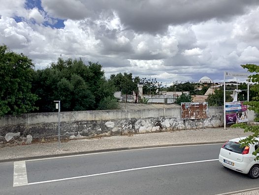
A couple of weeks ago our Portuguese correspondent Orwell sent in this photo of a billboard in Loulé in the Algarve celebrating the centennial of communism in Portugal:

At some point since then the red billboard, like so many enemies of communism, has been disappeared:

Orwell had this to say about the vanished billboard:
It’s bizarre!
It looks like someone else has taken solid action to remove the huge PCP communist banner ad. Not only that, but the frame supporting it has also been completely demolished.
Rightly so.
Note: For the title of this post I thought up “Red B Gon” as an imaginary (but much-needed) product that eliminates communism at home and in the workplace.
It’s my habitual practice to do an internet search for any clever idea I come up with, because 95% of the time someone has already thought of the same thing. In this case, a product called “Red-B-Gone” already exists. Its purpose is to get rust out of water pumps and similar devices where the constant presence of water causes rust to accumulate. However, I couldn’t find any product by the same name that is designed to get rid of commies, so the diligent householder will just have to do that particular job himself.
 The family of Andrew Brown Jr., who was fatally shot by deputies in Elizabeth City, North Carolina, said that Mr. Brown had his hands on the steering wheel when he was “executed” by police. Elizabeth City has declared a state of emergency.
In other news, a federal court in Canada has ruled that Justin Trudeau’s “COVID hotels” violate protections against arbitrary detention.
To see the headlines and the articles, click “Continue reading” below.
Thanks to Dean, DV, LP, Reader from Chicago, SS, and all the other tipsters who sent these in.
Notice to tipsters: Please don’t submit extensive excerpts from articles that have been posted behind a subscription firewall, or are otherwise under copyright protection.
Caveat: Articles in the news feed are posted “as is”. Gates of Vienna cannot vouch for the authenticity or accuracy of the contents of any individual item posted here. I check each entry to make sure it is relatively interesting, not patently offensive, and at least superficially plausible. The link to the original is included with each item’s title. Further research and verification are left to the reader.